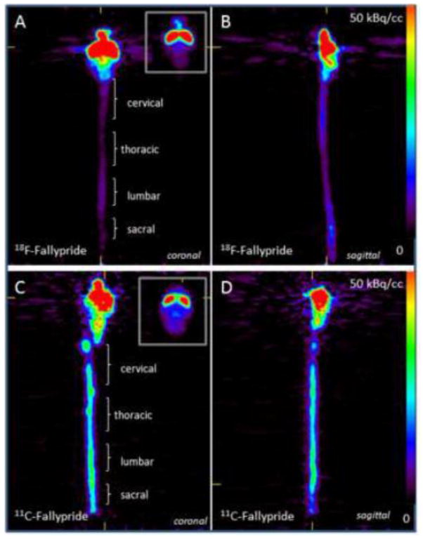
Figure 5. Rat Ex Vivo PET studies with 18F-Fallypride and 11C-Fallypride of the brain and spinal cord.
Ex vivo PET image of a rat sacrificed after initial PET scan (Figure 4) and the brain and spinal cord excised for ex vivo PET imaging (n=2). Ex vivo 18F-fallypride (A and B) and 11C-fallypride (C and D) images of brain and spinal cord in the coronal (A and C) and sagittal view (B and D) are shown. Binding of 18F-fallypride and 11C-fallypride is evident in the brain (striatum, at the top right corner of A and C) and in the various regions of the spinal cord as shown in C (cervical, thoracic, lumbar, sacral).

