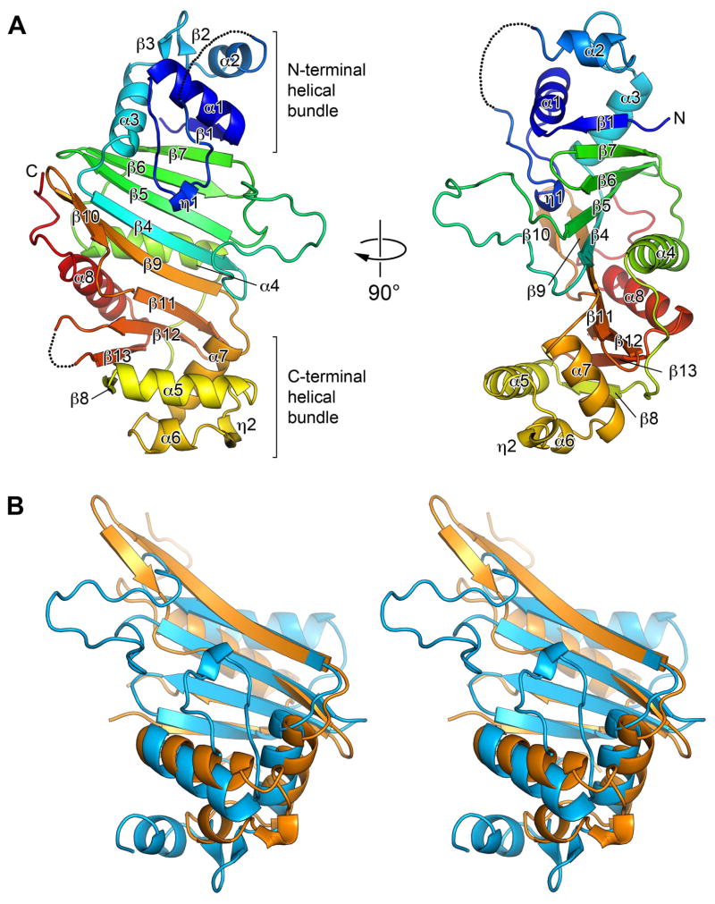
Fig. 2. Structure of EspG5 represents a novel fold with quasi 2-fold symmetry.
A. Ribbon representation is colored in rainbow colors from N-terminus (blue) to C-terminus (red). Secondary structure elements are labeled α1–α8 and β1–β13. The disordered loops are indicated as dashed lines. The two views are related by ~90° rotation.
B. Stereo view of the structural superposition of the N-terminal (blue) and the C-terminal (orange) sub-domains of EspG5.

