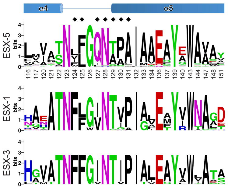
Fig. 5. EspG5-binding region of PPE41.
The sequence conservation of the EspG5-binding site is displayed as a sequence logo (http://weblogo.berkeley.edu) (Crooks et al., 2004) based on the sequence alignments of ESX-5- and ESX-3-specific PPE proteins of M. tuberculosis H37Rv and ESX-1-specific PPE68 homologs from mycobacteria (Fig. S2, S6 and S7). Secondary structure elements of PPE41 are shown at the top. Black diamonds indicate residues that were subjected to mutational analysis (Table 2 and Fig. 3C, S8, S9 and S10).

