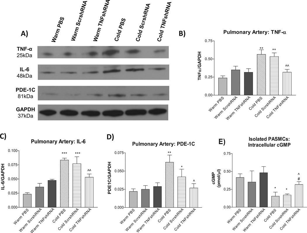
Figure 5. TNFshRNA prevented cold-induced increases in TNF-α, IL-6, and PDE-1C expression in pulmonary arteries (PAs).
Western blot was used to measure protein expression of pulmonary artery TNF-α, IL-6 and PDE-1C. A) Representative Western blots of TNF-α, IL-6 and PDE-1C protein expression. Quantitative analysis of B) TNF-α expression, C) IL-6 expression, and D) PDE-1C expression in PAs. E) TNFshRNA delivery increased intracellular cGMP in isolated PASMCs. A cGMP ELISA kit was used to measure intracellular cGMP in isolated PASMCs. Data=means±SEM. *p<0.05, **p<0.01, ***p<0.001 vs. Warm PBS; ^p<0.05, ^^p<0.01 vs. Cold PBS. #p<0.05 vs. Cold ScrshRNA. N=6.

