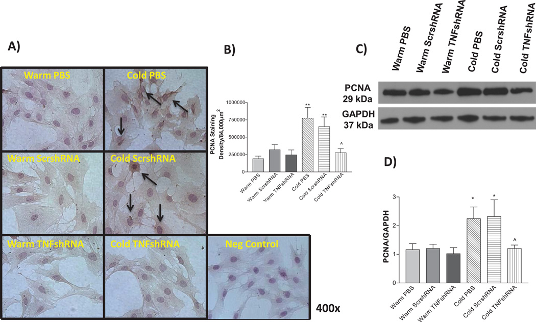
Figure 8. TNFshRNA prevented the cold-induced increase in PCNA expression.
A) IHC analysis using isolated PASMCs. Cells were stained with an anti-PCNA primary antibody. B) Densitometry analysis of the IHC PCNA staining in PASMCs. C) Representative Western blot of PCNA staining and D) Quantitative analysis of PCNA expression, as measured by Western blot. Photos are shown at 400×. Arrows indicate positive PCNA staining (brown). Data=means±SEM. *p<0.05, **p<0.01 vs. Warm PBS; ^p<0.05 vs. Cold PBS. N=6.

