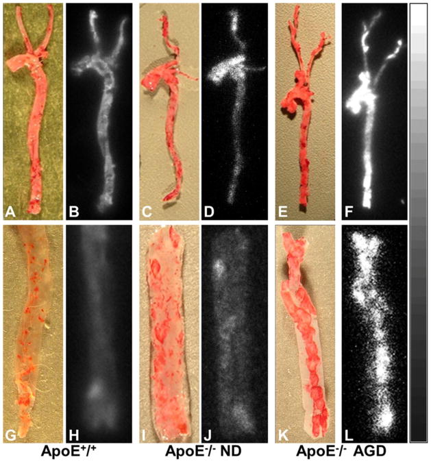
Fig. 5.
Photographs of Oil Red O staining vs. autoradiographs of intact mouse aortas (top row) and incised aortic segments (bottom row) from an ApoE+/+ mouse (A, B, G, and H), ApoE−/− mouse on ND (C, D, I, and J), and ApoE−/− mouse on AGD (E, F, K, and L) at age 48–52 weeks. Autoradiographs showed significantly higher radioactive uptake in aortic and carotid artery lesions stained by Oil Red O in the Apo-E−/− mouse on AGD (F, L), but barely detectable radioactive accumulations in the corresponding vessels of the ApoE+/+ mouse (B, H). The ApoE−/− mouse on ND exhibited slightly increased uptake in the aorta and carotid artery (D, J).

