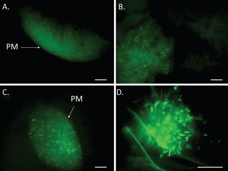
Figure 2. GFP-Escherichia coli O157:H7 in the alimentary canal of the house fly.

House flies (total n = 28 in three biological replicates) were fed 1.8 ± 0.9 × 10⁶ CFU (mean ± SE) GFP-expressing E. coli O157:H7. Bacterial cells (green rods) were visible in the midgut at all time points including 2 h PI (A), 4 h PI (B), and 6 h PI (C). Clumps of intact cells were visible in the crop at all time points including 12 h PI (D) in one replicate. PM, peritrophic matrix. Scale bar is 10 μm.
