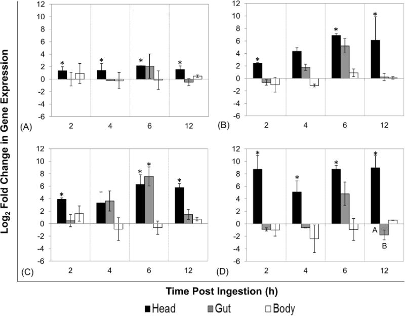
Figure 3. Expression of antimicrobial peptides and lysozyme in house flies after ingestion of GFP-Escherichia coli O157:H7.

Flies (total n = 95 in three biological replicates) were fed GFP-expressing E. coli O157:H7 (mean CFU ± SE: 1.40 ± 0.30 × 10⁶) and aseptically dissected at 2, 4, 6, and 12 h post-ingestion (n=7–8 per time point) to separate heads (with salivary glands), guts (i.e. proventriculus to rectum), and carcasses. qRT-PCR analysis was used to determine relative tissue-specific expression of cecropin (A), defensin (B), diptericin (C), and lysozyme (D) in house fly tissues (pooled per tissue within replicate). Log2 fold change in expression was determined using the REST-MCS® software, with calibration to matched tissues from broth-fed control flies and the rps 18 reference gene. The mean expression of three biological replicates along with standard error is shown. Different letters denote statistical significance (P < 0.04) between tissues within time point. Asterisks denote statistical significance from broth-fed control flies (P < 0.05)
