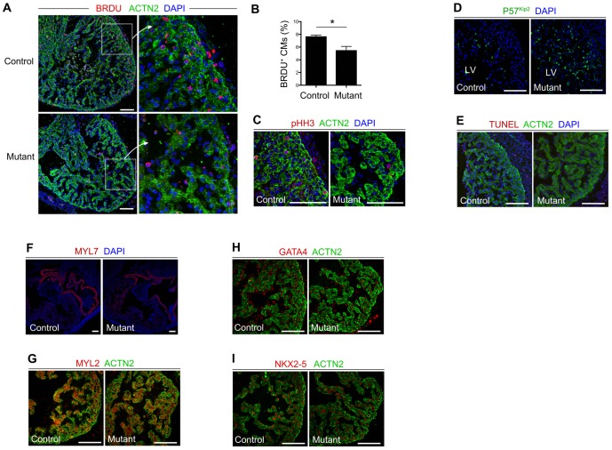
Figure 3. Reduced cardiomyocyte proliferation in BAF200 mutant hearts.
(A, B) Staining of BRDU shows reduced cardiomyocytes (CMs) proliferation in E13.5 BAF200 mutant hearts compared with littermate controls. *P<0.05. (C) phospho-histone H3 staining shows reduced proliferation in BAF200 mutant compact myocardium. (D) Expression of p57kip2, a cyclin-dependent kinase inhibitor, is increased in mutant hearts. (E) No significant apoptosis was detected in mutant and control embryos. (F, G) Expression of chamber-specific markers MYL2 and MYL7 was unchanged in BAF200-deficient hearts. (H, I) Expression of cardiac transcription factors GATA4 and Nkx2-5 were not significantly changed in BAF200 mutants. White bar = 100 um.

