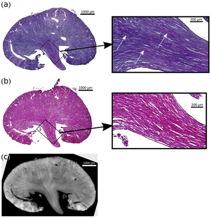
Figure 7. Comparison of Histological Slices of Clamped Kidney.
(a) PAS histological slice of the clamped kidney. (b) HE-stained histological slice of the clamped kidney, with a similar slice of the phase-contrast volume (c). For both histology slices a respective zoom view of the inner medulla is given. Scarring and beginning tubular atrophy as well proteinaceous casts could be detected correlating with increasing medullary density in PCI.

