
Fig. 3.
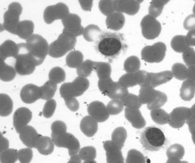
Peripheral blood—hairy cell with round nucleus, homogenous spongy chromatin with abundant pale blue cytoplasm with circumferential hair like projections. (Jenner Giemsa stain, ×1,000). (Color figure online)
Official websites use .gov
A
.gov website belongs to an official
government organization in the United States.
Secure .gov websites use HTTPS
A lock (
) or https:// means you've safely
connected to the .gov website. Share sensitive
information only on official, secure websites.
Peripheral blood—hairy cell with round nucleus, homogenous spongy chromatin with abundant pale blue cytoplasm with circumferential hair like projections. (Jenner Giemsa stain, ×1,000). (Color figure online)