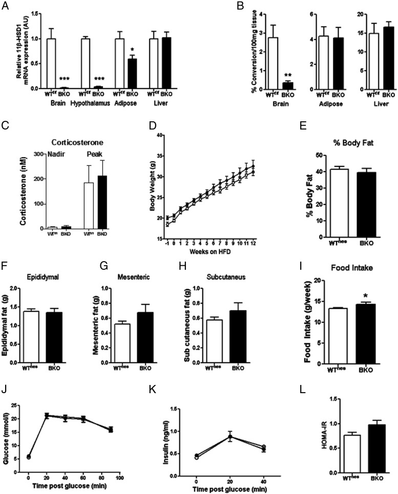
Figure 2.
BKO mice have no reduction in body weight, fat mass, food intake, or glucose tolerance. A, 11β-HSD1 mRNA expression in brain, hypothalamus, epididymal adipose, and liver tissues. B, Enzyme activity in brain, epididymal adipose tissue, and liver in BKO and WTf/f mice. C, Circulating corticosterone concentrations in BKO and WTf/f mice after 18 weeks of HFD feeding. D, Body weight over 12 weeks of HFD feeding in BKO (■) and WTnes (□) mice. E, Percentage body fat measured by DEXA scan after 12 weeks of HFD feeding. F–H, Epididymal (F), mesenteric (G), and subcutaneous (H) fat pad masses after 14 weeks of HFD feeding. I, Average food intake (grams per week) over 12 weeks of HFD feeding. J and K, Glucose (J) and insulin (K) excursion during an OGTT after 13 weeks of HFD feeding. L, HOMA-IR after 13 weeks of HFD feeding. Data are expressed as means ± SEM; n = 6 to 16. *, P < .05; **, P < .01; ***, P < .001 vs the appropriate WT strain.

