
Figure 1.
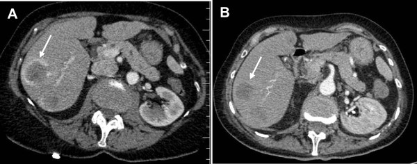
Computed tomography. The figures shows the initial CT scan of the liver (performed at the time that the metastatic stage of the disease was diagnosed) (A) and a CT scan acquired 4 weeks after the initiation of vemurafenib treatment (B). The metastasis is indicated by an arrow. Comparison of the two scans reveals a partial regression of the metastatic tumor.