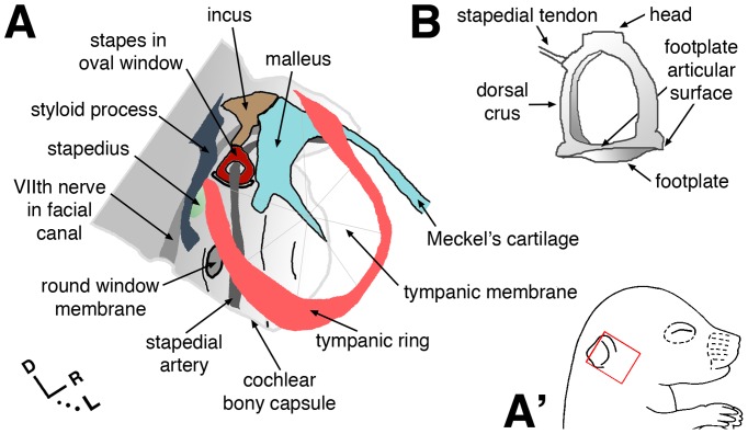
Figure 1. Schematic views of selected middle ear structures and otic capsule.
(A) Anatomy conforms to that of an excised mouse temporal bone at post-natal day 5, viewed laterally. Orientation within the head is indicated by the red box in A′, bottom right. R in axes = rostral. (B) Caudal view of the isolated adult stapes.

