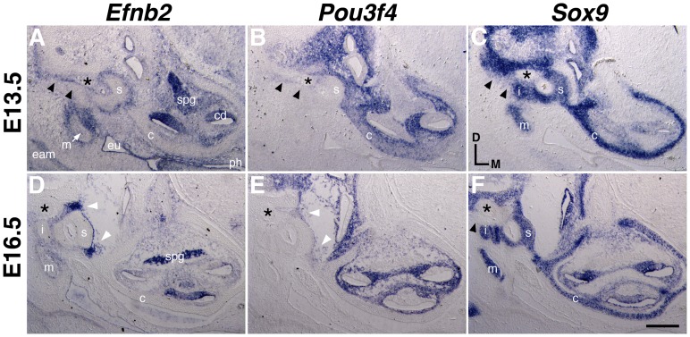
Figure 4. Comparative expression of Efnb2, Pou3f4, and Sox9 at E13.5 and E16.5.
Adjacent transverse sections through embryos at two developmental stages, hybridized to detect Efnb2 (A,D), Pou3f4 (B, E), or Sox9 (C, F). c, cartilaginous cochlear capsule; cd, cochlear duct; eam, external auditory canal; eu, nascent eustacian tube; i, incus; m, malleus; ph, pharynx, spg, spiral ganglion, s, stapes. Asterisk highlights the facial nerve. Double arrowheads in (A–C) highlight overlapping Efnb2 and Pou3f4 signals and relatively weak Sox9 signal dorsal to the stapes. Single arrowhead in (F) highlights a lack of Sox9 signal in the corresponding region at E16.5. White arrowheads in (D, E) highlight overlapping Efnb2 and Pou3f4 signals at the forming S–V joint. Scale bar represents 200 microns in (A–C) and 250 microns in (D–F).

