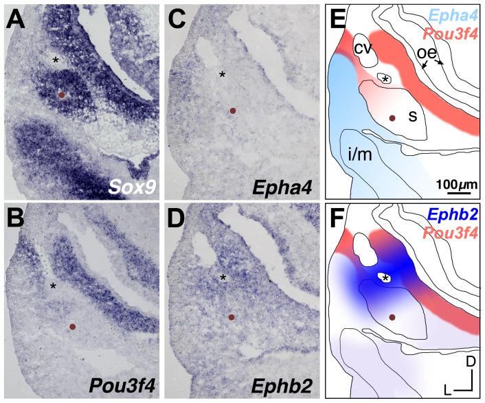
Figure 6. Comparative expression of Sox9, Pou3f4, Epha4, and Ephb2 at E11.5.
(A–D) Adjacent transverse sections through a wild-type E11.5 embryo, hybridized to detect Sox9, Pou3f4, Epha4, or Ephb2. Mesenchyme that will form the stapes condensation is identified by Sox9 signal surrounding the stapedial artery (brown dots) and is located ventral to the facial nerve (asterisks). (E, F) Schematized spatial relationships between expression of Pou3f4 and either Epha4 (E) or Ephb2 (F). Asterisks denote VIIth nerve; brown dots denote the stapedial artery. cv, cardinal vein; oe, otic epithelium; s, stapes pre-condensation; i/m, incus/malleus pre-condensation. All panels are shown to scale.

