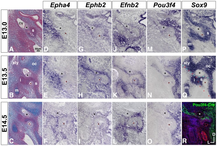
Figure 7. Comparative developmental expression of Epha4, Ephb2, Efnb2, Pou3f4, and Sox9 at the stapes condensation and neighboring structures.
(A–C) Transverse histological sections through wild-type embryos at E13, E13.5, and E14.5, stained with alcian blue/nuclear fast red. Asterisks highlight the VIIth nerve; brown dots or open arrow in (A) highlight the stapedial artery. cv, cardinal vein; s, stapes condensation; sty, styloid process condensation; oc, otic capsule cartilage; i, incus condensation; m, malleus cartilage. (D–Q) Developmental gene expression in wild-type embryos. Annotations are as in (A–C). Red and cyan dotted lines in (Q) highlight salient borders of Epha4 and Ephb2/Efnb2/Pou3f4 expression, respectively. (R) Pou3f4-Cre-mediated ROSA-YFP reporter expression at E14.5. Cre-positive cells or descendents of Cre-positive cells are labeled green. Section is counterstained with phalloidin (red) and DAPI (blue). All photos are shown to scale.

