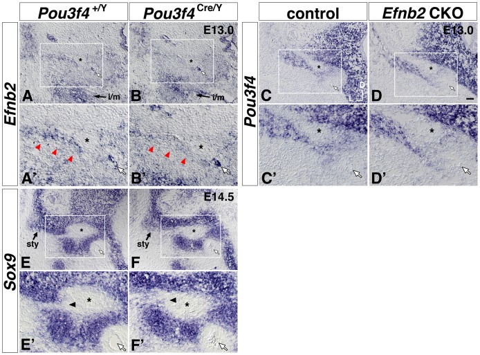
Figure 8. Attenuated Efnb2 signal and altered patterning of Sox9 signal near the Pou3f4 Cre/Y stapes condensation, but no apparent change of Pou3f4 signal in the Efnb2 CKO.
(A, A′, B, B′) Transverse sections of control (A, A′) and Pou3f4 Cre/Y mutant (B, B′) E13.0 littermates hybridized to detect Efnb2. Red arrowheads in (A′, B′) highlight the medial-lateral coursing stripe of Efnb2 signal dorsal to the stapes condensation. i/m, Efnb2 signal at the incus and malleus rudiment. (C, C′, D, D′) Transverse sections of control (C, C′) and Sox9-IRES-Cre-mediated Efnb2 CKO (D, D′) E13.0 littermates hybridized to detect Pou3f4. (E, E′, F, F′) Transverse sections of control (E, E′) and Pou3f4 Cre/Y mutant (F, F′) E14.5 littermates hybridized to detect Sox9. Arrowheads in (E′, F′) highlight an alteration in Sox9 patterning across genotypes. sty, styloid process condensation. Boxed regions of interest highlighting the stapes and surrounding structures in (A–F) are shown at 2x magnification in (A′–F′), respectively. Scale bar in (D) = 50 micron for (A–F), and 25 microns for (A′–F′). In all panels, white arrows highlight the stapedial artery, and asterisks highlight the VIIIth nerve. Axes in (C) apply to all photos.

