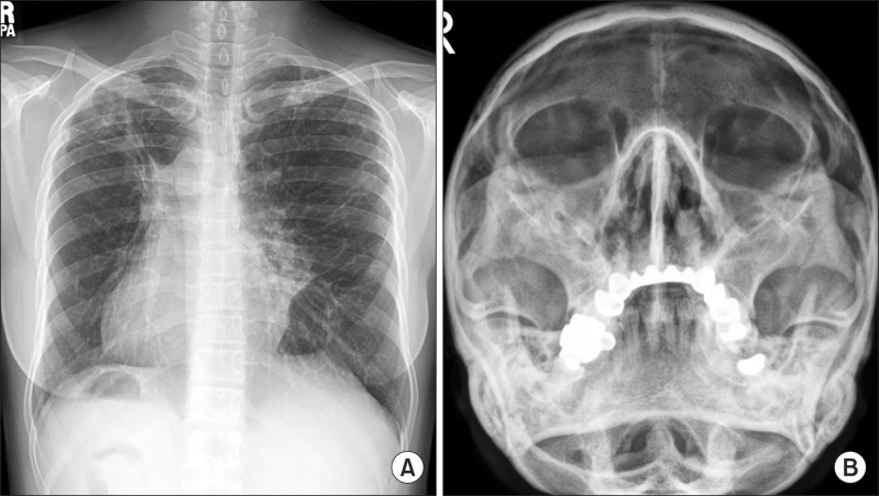
Figure 1.
(A) Chest radiography showed dextrocardia and situs inversus. Note the cavitary lesions in the right upper lobe and nodulostreaky opacity, suggesting bronchiectasis in the left middle lung zones. (B) Paranasal sinus radiography showed total opacification involving the bilateral ethmoid sinus and bilateral maxillary sinus.

