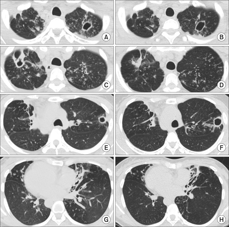
Figure 2.
(A, C, E, G) Chest high-resolution computed tomography showed multiple cavities in both upper lobes. Note the severe bronchiectasis in the left middle lobe and the lingular segment of the right upper lobe. (B, D, F, H) Although the cavitary lesion in the right upper lobe improved after 20 months of antibiotic treatment, the size of the multiple cavities in the left upper lobe increased.

