
FIGURE 8.
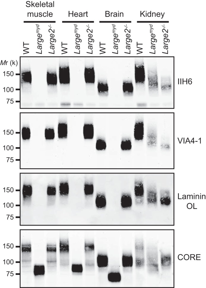
Functional glycosylation of α-DG in tissues of the WT, Largemyd, and Large2−/− mice. Immunoblotting or laminin overlay (OL) of WGA-enriched glycoproteins extracted from tissues of the WT, Largemyd, or Large2−/− mouse. The α-DG antibodies used are: IIH6, VIA4–1 (recognize glycan epitopes) and CORE (recognizes a core protein epitope). Mr, relative molecular mass.