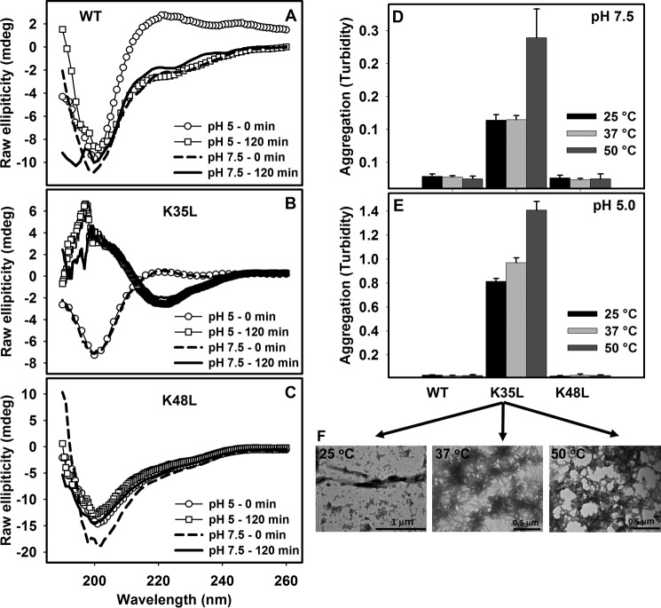
FIGURE 4.
Secondary structure determination and aggregation properties of WT, K35L, and K48L 26–57 peptides. CD spectra of the WT (A), K35L (B), or K48L (C) peptides immediately after dilution in buffers pH 5.0 (circles) or pH 7.5 (dashed lines). The peptides were allowed to aggregate at 25 °C for 120 min at pH 5 (squares) and pH 7.5 (continuous line), and their CD spectra were recorded. Shown are end points of aggregation reactions of the three peptides at 100 μm for 12 h at pH 7.5 (D) or pH 5.0 (E) at 25 °C (black bar), 37 °C (light gray bar), and 50 °C (dark gray bar) as measured by absorbance at 330 nm. F, TEM images of the aggregates of K35L formed at pH 5.0 at 25, 37, and 50 °C (from left to right, respectively). Bars, 1 or 0.5 μm, as indicated. Error bars, S.E.

