
FIGURE 3.
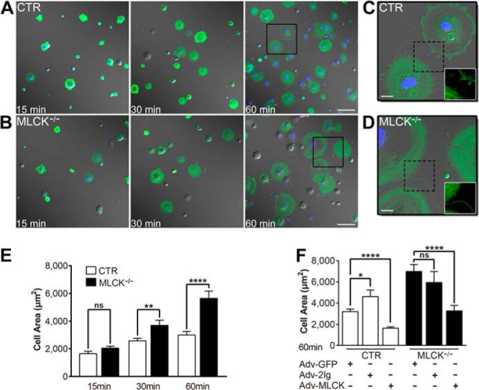
MLCK-deficient SMCs show accelerated protruding and spreading in vitro. A and B, the spreading assay for the SMCs was performed by seeding the suspended SMCs on gelatin-coated coverslips followed by imaging at 15, 30, and 60 min. The SMCs were visualized with an anti-SMA antibody (green) and TO-PRO-3 (blue). DIC images merged with fluorescence signals are shown. C and D, magnified images derived from the boxed areas in panels A and B, respectively. Fluorescent images of dashed boxed areas are shown in white boxes. E, the area of spreading cells was measured (n = 46–89) and statistically analyzed. F, rescue assay for MLCK-deficient SMCs. Control and MLCK-deficient SMCs were infected with control adenovirus (Adv-GFP) and MLCK-expressible variants (Adv-MLCK, Adv-2Ig; MOI = 4) for 48 h, and then the 60-min spreading area was examined. n = 32–88. Scale bars are 100 μm (A, B) and 20 μm (C, D).