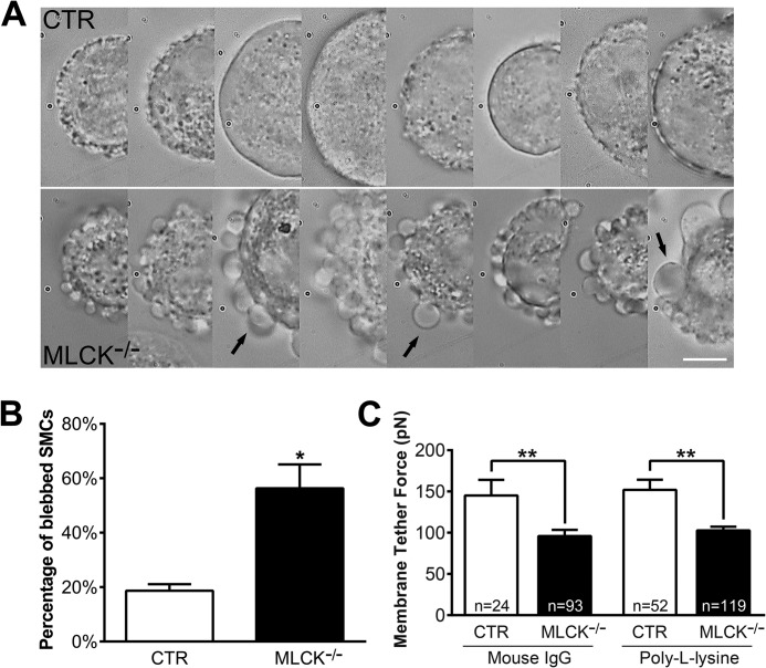
FIGURE 4.
Membrane tether force is reduced in MLCK-deficient SMCs. The SMCs cultured in vitro were isolated by TrypLETM Express digestion and then subjected to tether force measurement with laser tweezers. A, DIC images show many membrane blebs around MLCK-deficient SMCs (below) after suspending over 20 min, but very few were observed around controls (above). Arrows point to typical membrane blebs. The scale bar is 10 μm. B, percentages of the blebbed cells were calculated (CTR: n = 302; KO: n = 193). C, average membrane tether forces measured by laser tweezers with mouse IgG- and poly-l-lysine-coated microbeads.

