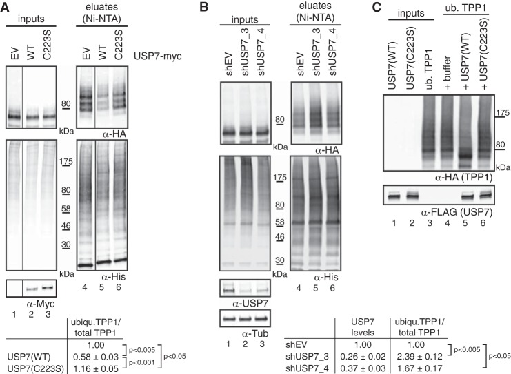
FIGURE 3.
TPP1 is deubiquitinated by USP7. A, cells were co-transfected with His-ubiquitin, TPP1–3xHA, and the indicated USP7 constructs. 48 h post-transfection, cells were harvested followed by urea extraction (lanes 1–3), Ni-NTA binding (lanes 4–6), and Western blot analysis. Vertical lines separate different parts of the same gel. Quantification of ubiquitinated (ubiqu.) TPP1 over total TPP1 from three independent experiments reveals that USP7(WT) but not inactive USP7(C223S) reduces TPP1–3xHA ubiquitination. Average values, standard deviations, and p values as determined by t test are indicated. B, cells were transfected with the indicated shRNA constructs. Puromycin selection was started 20 h post-transfection and continued throughout the experiment. 48 h after shRNA transfections, cells were transfected with His-ubiquitin and TPP1–3xHA. After a further 48 h, cells were harvested followed by urea extraction (lanes 1–3), Ni-NTA binding (lanes 4–6), and Western blot analysis. Quantification from three independent experiments shows that USP7 depletion increases TPP1–3xHA ubiquitination. Average values, standard deviations, and p values as determined by t tests are indicated. C, ubiquitinated (ub.) TPP1 was purified as described under “Experimental Procedures” (lane 3) and incubated with either buffer (lane 4), purified USP7(WT) (lane 5), or USP7(C223S) (lane 6). Western blot analysis reveals that USP7(WT) but not USP7(C223S) deubiquitinates TPP1–3xHA in vitro. EV, empty vector; Tub, tubulin.

