
FIGURE 8.
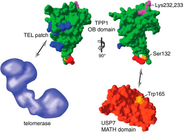
Structural representation of the TPP1(OB) domain interactions and ubiquitination sites. The structure of the TPP1(OB) domain is shown in green (Protein Data Bank code 2I46) with the TEL patch region in blue, the USP7 consensus motif in red (Ser-132 in orange), and the ubiquitination sites Lys-232 and Lys-233 in magenta. The structure of the MATH domain of USP7 is shown in red (Protein Data Bank code 1YZE) with Trp-165 in orange. Structure images were generated with Swiss-Pdb Viewer (38). The schematic representation of telomerase in blue is not drawn to scale. The TPP1(OB) domain interacts with telomerase and USP7 via distinct surfaces. Although the ubiquitination sites of the TPP1(OB) domain appear to be on a different surface than the USP7 consensus motif, they are likely accessible for the catalytic peptidase domain of USP7, allowing USP7 to deubiquitinate TPP1.