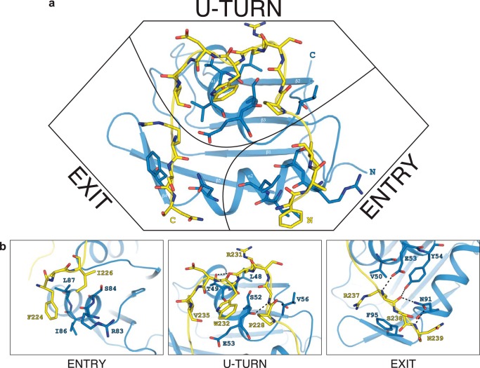
FIGURE 7.
Details of the intermolecular contacts in the Snu17p-Bud13p complex. a, the Bud13p peptide forms an arch around the helical binding surface of Snu17p. b, close-up views of the interaction at the entry point (left), the U-turn (middle), and the exit point (right) of the Bud13p peptide shown in a. Interacting residues are annotated and colored blue for Snu17p and yellow for Bud13p. Intermolecular hydrogen bonds to the Bud13p backbone are indicated by dashed lines.

