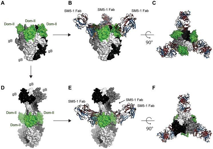
Figure 7. SM5-1 Fab is able to bind to both a prefusion and a postfusion model of entire trimeric HCMV gB.
(A) Prefusion conformation of trimeric HCMV gB modelled according to the crystal structure of VSV-G (PDB ID 2j6j, [19], [26]). (B and C) Two orthogonal views of SM5-1 Fab bound to the prefusion conformation of gB. (D) Postfusion conformation of HCMV gB modelled according to the crystal structure of HSV-1 gB (PDB ID 2gum, [18]). (E and F) SM5-1 bound to the postfusion conformation of gB. The arrow linking panel A to C indicates the conformational transition from a prefusion to a postfusion model of HCMV gB. The arrows linking panel A and B as well as D and E represent SM5-1 binding. Trimeric gB is shown in a surface representation and colored white, grey and black with gB Dom-II highlighted in green. SM5-1 Fab is shown in a cartoon representation.

