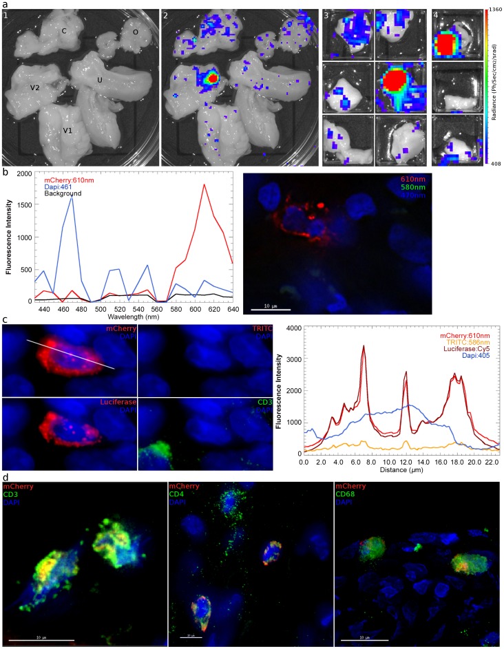
Figure 4. Identification and phenotyping of infected cells from female reproductive tract. a,
48 hours after inoculation with JRFL pseudotyped LICh vector, the reproductive tract is separated into large sections (panel 1; V1: labia and lower vagina; V2: upper vagina; C: cervix; U: uterus; O: ovary), soaked in d-luciferin to identify luciferase expressing regions with in vivo imaging systems, and dissected into sequentially smaller pieces (panels 2–4) to isolate strongly luminescent regions measuring 2×2 mm2. (animal code: EH99) b, Left panel; The spectral emission profile of a putative infected cell from the tissue identified in a is measured on laser scanning confocal microscope with resonant scanner. Emission profile matches the defined profile of mCherry. Right panel; Pseudo-colored reconstructed image on the right shows the localization of mCherry (red), DAPI (blue) and background signals (green). c, Vaginal tissue is stained for firefly luciferase expression. The combination of mCherry+ (red), TRITC−/low (red), luciferase+ (red) fluorescence is criteria for confirming infection. The right panel shows relative fluorescence intensity of mCherry, TRITC, luciferase, and nucleus is along the line in mCherry image. (animal code: HP79) d, Phenotyping of infected cells by surface marker staining, shown in green, for CD3 (T Cell), CD4 (HIV-1 primary receptor), or CD68 (tissue resident macrophages) shows all conventionally defined HIV susceptible cell types are transduced within the vaginal tissue shown in a. (animal code: EH99).

