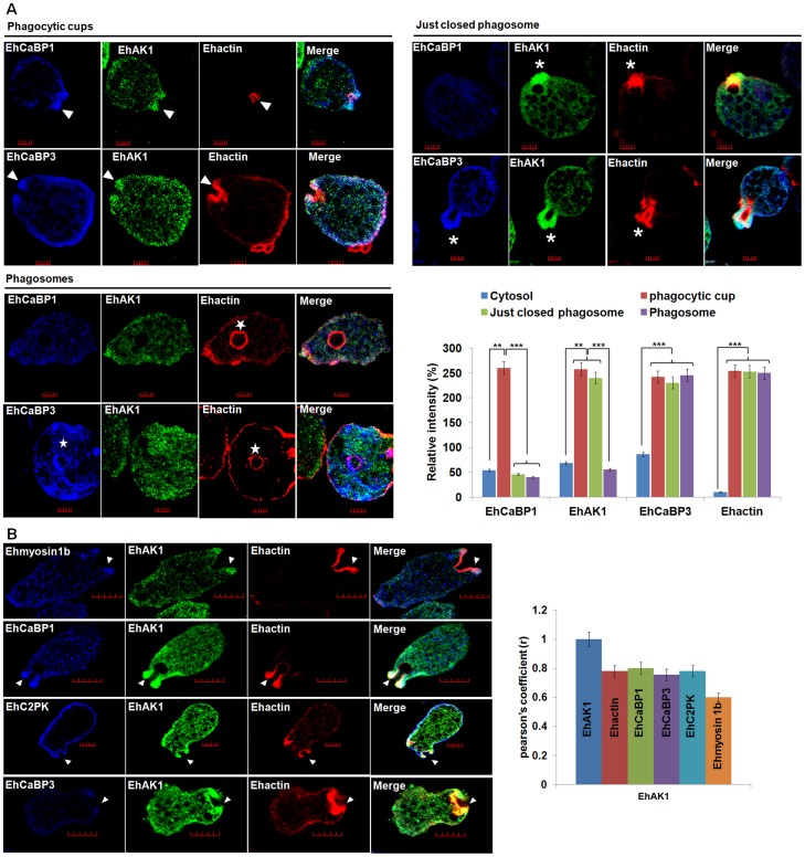
Figure 2. Colocalization of EhAK1 with EhMyosin 1B, EhC2PK, EhCaBP1 and EhCaBP3 at the phagocytic cup during erythrophagocytosis.
(A) Imaging of EhAK1, EhCaBP1 and EhCaBP3 during erythrophagocytosis. E. histolytica cells were incubated with RBC for 5 min at 37°C. The cells were then fixed and immunostained with anti-EhAK1 antibody followed by Alexa-488. F-actin was stained with TRITC-phalloidin and other indicated proteins were immunostained with respective antibodies and followed by Pacific blue-410. Arrowheads indicate phagocytic cups, asterisks mark just closed cups and star marks phagosome. Bar represents 5 µm. Quantitative analysis of fluorescent signals obtained by immunostaining of EhAK1, EhCaBP1 and EhCaBP3 from different locations in E. histolytica cells (N = 5) was done as described in Fig. 1D. (B) The incubation and labelling conditions were as described in Fig. 1. Ehmyosin 1B, EhC2PK, EhCaBP1 and EhCaBP3 were immunostained with specific antibodies and visualized using Pacific blue-410 (blue). Colocalization analysis from five cells was done by using JACoP (ImageJ). The Pearson's coefficient (r) of EhAK1 with EhAK1, EhCaBP1, EhCaBP3, Ehmyosin 1B and EhC2PK from phagocytic cups are indicated. *p-value≤0.05, **p-value≤0.005, ***p-value≤0.0005.

