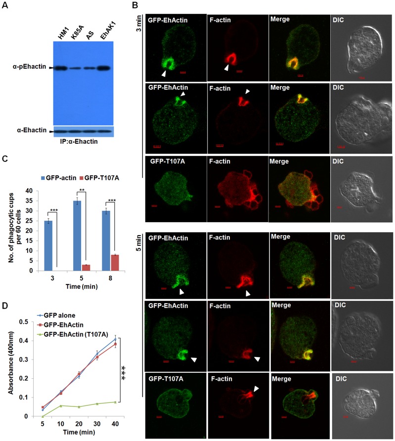
Figure 8. Phosphorylated actin participates in phagocytosis.
(A) E. histolytica cells carrying EhAK1 construct in the sense and the antisense orientations and K85A in the sense orientation as indicated were induced with tet. Phosphorylated actin was detected by immunoprecipitation using anti Ehactin antibody followed by western blots using anti-pEhactin antibody. (B) Indicated cell lines grown with 30 µg/ml G418 to enhance the expression of transfected gene were incubated with RBC for 3 min at 37°C. The cells were then fixed and immunostained with anti-GFP. F-actin was stained with TRITC-phalloidin. Arrowheads indicate phagocytic cups. Bar represents 20pixels. A few representative cells are shown. (C) Quantitative analysis was carried out by selecting randomly sixty cells from each experiment and the numbers of phagocytic cups present in all cells were counted (blue, GFP-actin; red, GFP-T107A). (D) RBC uptake assay was performed using indicated cells grown with G418. The experiments were carried out three times independently in triplicates. *p-value≤0.05, **p-value≤0.005, ***p-value≤0.0005.

