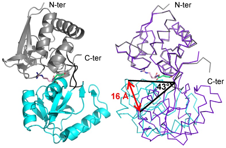
Figure 1. Ribbon representation of NocT in complex with nopaline and comparison with the unliganded form.
Left panel; Nopaline is located in the cleft between the two domains shown in grey and cyan. The short hinge region between the two domains is shown in black. The arginine part of the nopaline is shown in pink while its α-KG part in limegreen. Right panel, comparison of the unliganded structure of NocT in purple with the complexed nopaline in grey and cyan.

