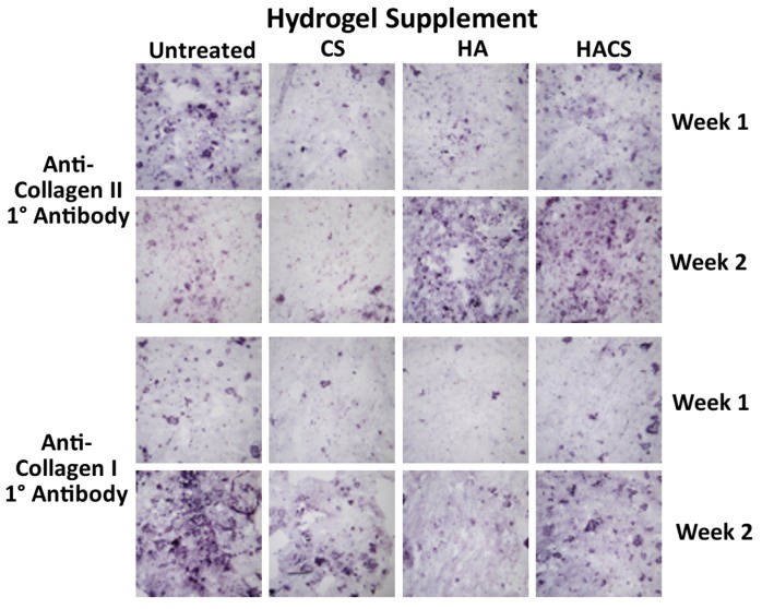
Figure 4.
Immunostaining of cryogenic sections of hydrogels supplemented with CS, HA or HACS. Non-supplemented hydrogels (untreated) served as controls. Staining revealed that there was an accumulation of collagen types I and II proteins over time in all treatment groups. All photomicrographs were taken at equivalent magnification on a Leitz DM-RB compound microscope (10× objective lens) equipped with a Sony DSC-V3 camera.

