
Figure 2. False-positive fluorescence readings (620 nm excitation/680 nm emission) from biological solutes.
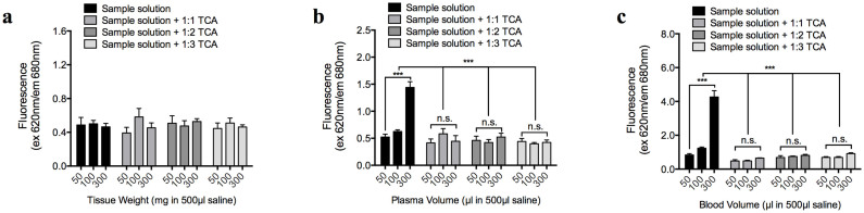
(a–c) Auto-fluorescence of biological solutes from the brain (a), blood (b), and plasma (c) samples (diluted to 500 µl in saline), with or without protein-precipitation by 1:1–1:3 50% trichloroacetic acid (TCA). [TCA final] following the addition of 1:1, 1:2, and 1:3 volume ratios of 50% TCA were 25, 33, and 37.5%, respectively.