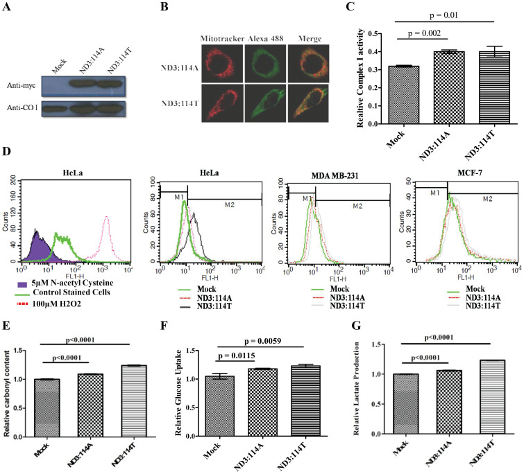
Figure 1.
(A) Western blots (Cropped) with anti-myc antibody showing the expression of exogenously expressed ND3:114A and ND3:114T; Anti-COI western shows loading control (Full length blots are provided in Supplementary figure 4); (B) Fluorescence confirming mitochondrial localization of exogenously expressed protein, mitochondria were stained with 25 nM mitotracker, exogenously expressed protein were probed using alexa 488 linked secondary antibody for myc tag; (C) Enzyme activity of mitochondria enriched fraction for complex I from cells transfected with empty vector, ND3:114A and ND3: 114T; (D) Reactive oxygen species in mock, ND3:114A and ND3: 114T transfected cells. HeLa cells treated with 50 μM H2O2 and 5 μM N-acetyl cysteine treated cells were used as positive and negative control, respectively; (E) Relative carbonyl content; (F) Relative glucose uptake; and (G) Relative Lactate production.

