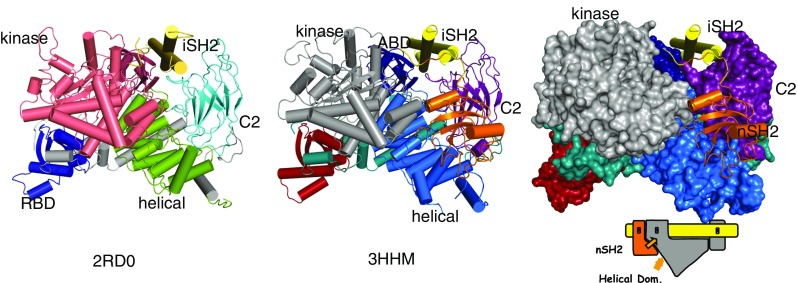
Fig. 1.

Structure of PI3Kα. a Structure of wild-type p110α/ni p85α heterodimer (pdb id 2RD0); the nSH2 domain is not shown because the experimental electron density for this domain was weak and diffuse. b Structure of the p110α/ni p85α heterodimer of the hot spot mutant H1047R showing the nSH2 domain. c Surface representation of the H1047R p110α/ni p85α with the kinase, C2 domains and helical in contact with nSH2
