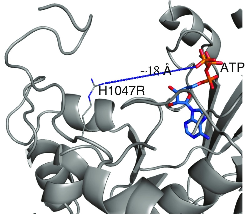
Fig. 3.
Position of the H1047R mutation in the p110α/ni p85αheterodimer structure. The top of the figure corresponds to the position of the membrane surface. The position of ATP is derived from the structure 1e8x (Walker et al. 1999). The kinase domain is colored gray and the other domains have been omitted

