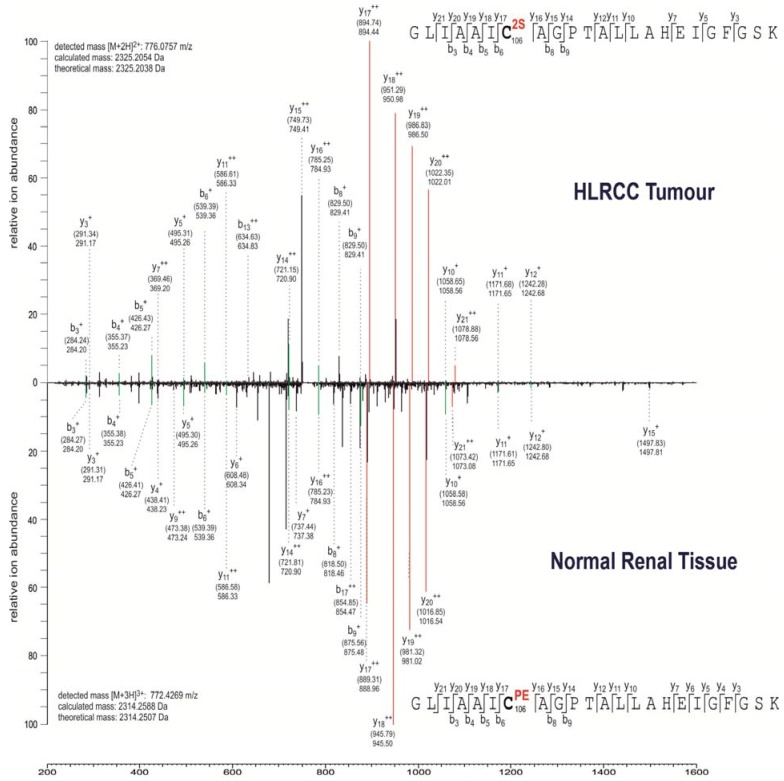
Figure 2.
MS/MS spectra showing succination of the active site (C106) of PARK7/DJ-1.
MS/MS spectra showing either succination (2SC) or pyridylethylation (PEC) at cysteine 106 in the 100GLIAAICAGPTALLAHEIGFGSK122 peptide of human DJ-1 derived from human HLRCC tumour tissue and normal renal tissue, respectively. Fragment ions are indicated for the peptide sequence and are labelled as follows: b: N-terminal fragment ion; y: C-terminal fragment ion; +: singly charged fragment ion and ++: doubly charged fragment ion. Both theoretical mass and detected mass (in brackets) are given for each assigned fragment ion. Matching fragment ion peaks between the two peptide species that do not contain the modified residue are highlighted in green, whereas peptide fragments of different mass that contain the modified residue are highlighted in red.

