
Figure 1.
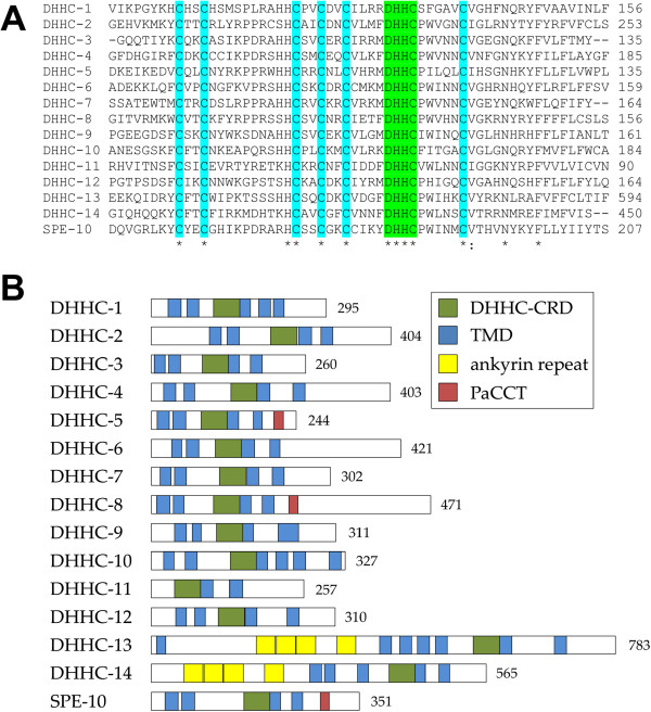
Sequence analysis of the putative C. elegans DHHC enzymes. (A) An alignment of the cysteine-rich domain (DHHC-CRD) of the putative C. elegans DHHC enzymes. The absolutely conserved DHHC motif is highlighted in green and conserved cysteine residues in cyan. The position of the final residue in the sequence shown is indicated on the right. (B) The predicted domain structure of the C. elegans DHHCs is represented in schematics drawn to scale. Features indicated are the DHHC-CRD (green), transmembrane domains (blue), ankyrin repeats (yellow) and palmitoyltransferase conserved C-terminal (PaCCT) (red). The sequence length in amino acids is indicated on the right.