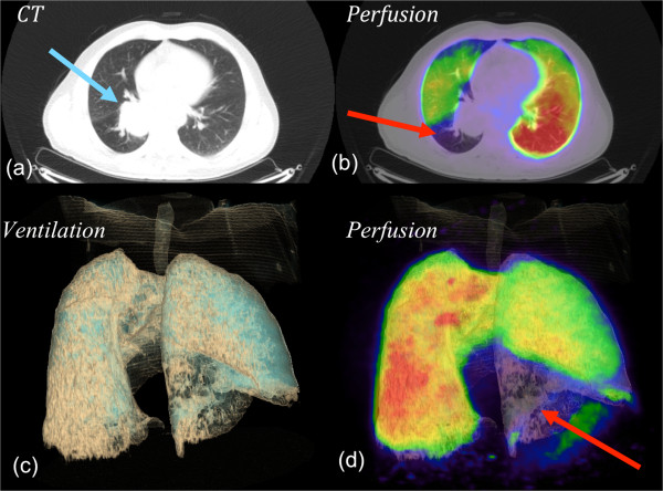
Figure 2.

V/Q PET/CT, a) CT alone, b) fused PET/CT, c) 3D volume rendered (VR) CT ventilation reconstruction d) 3D fused VR perfusion PET/CT. A patient with a large upper lobe NSCLC ( image a ), showing both ventilation deficits distal to tumour and perfusion deficits distal to the tumour (images b, c, and d).
