Fig. 1.
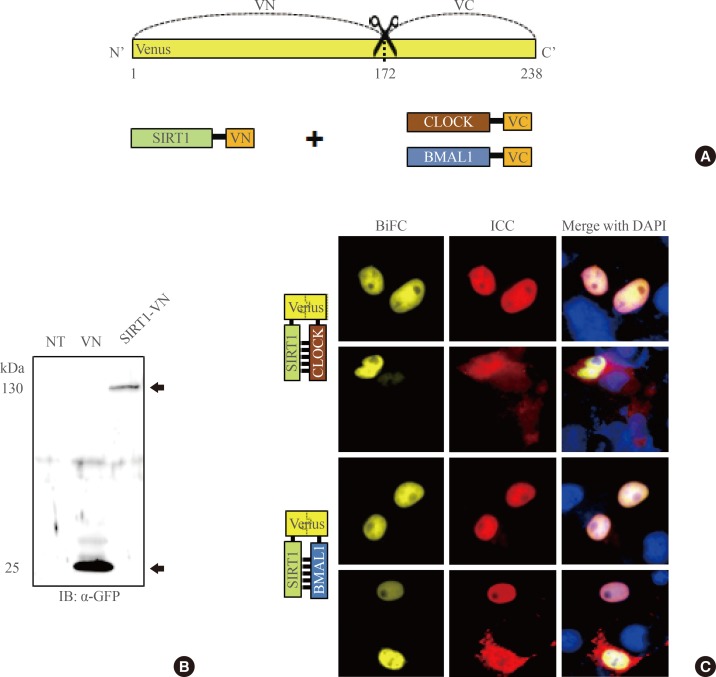
SIRTUIN1 interacts with CLOCK and BMAL1 in cell nuclei. (A) Schematic diagram of Venus-based bimolecular fluorescence complementation (BiFC) constructs. (B) Expression of SIRT1-N-terminal of Venus (VN) was detected by sodium dodecyl sulfate polyacrylamide gel electrophoresis separation followed by immunoblotting with an anti-green fluorescent protein (GFP) antibody (arrows). (C) Results from BiFC analysis and immunocytochemical experiments (ICC). COS7 cells were coexpressed with SIRT1-VN and either CLOCK-C-terminal of Venus (VC) or BMAL1-VC. Cells were immunostained with anti-GFP, anti-CLOCK, and anti-BMAL1 antibodies (red) to detect SIRT1-VN, CLOCK-VC, and BMAL1-VC, respectively. Nuclei were visualized with 4',6-diamidino-2-phenylindole (DAPI; blue). The images were acquired by fluorescence microscopy using specific filter sets for yellow fluorescent protein and red fluorescent protein. Scale bar=10 µm.

