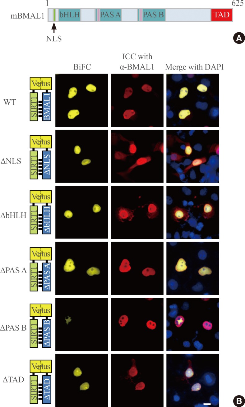
Fig. 2.
Identification of the SIRTUIN1 (SIRT1)-binding domain of BMAL1. (A) Schematic diagram of mouse BMAL1. (B) Binding of SIRT1 to BMAL1 deletion mutants was analyzed by bimolecular fluorescence complementation (BiFC). COS7 cells were transfected with plasmids encoding SIRT1-N-terminal of Venus (VN) and BMAL1-C-terminal of Venus (VC) wildtype (WT) or its various mutants (Δnuclear localization sequence [NLS], BMAL1 without a functional nuclear localization signal; Δbasic-helix-loop-helix [bHLH], encoding BMAL1 Δ71 to 140; ΔPer-Arnt-Sim [PAS]-A, BMAL1 Δ210 to 320; ΔPAS-B, BMAL1 Δ350 to 480; Δtranscriptional activation domain [TAD], BMAL1 Δ553 to 625). The images were captured by fluorescence imaging microscopy using specific filter sets for yellow fluorescent protein and red fluorescent protein. Scale bar=10 µm. ICC, immunocytochemical; DAPI, 4',6-diamidino-2-phenylindole.

