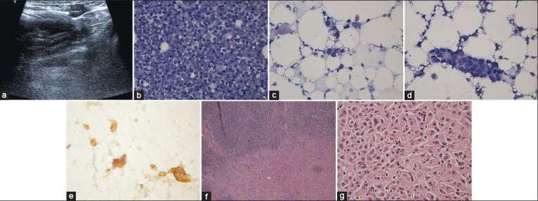
Figure 1.

Case with false negative fine needle aspiration cytology of axillary lymph nodes, ultrasound image (a). Smear 1 from case showing lymphoid cells only (Giemsa, ×400) (b). Smear 2 from case showing groups of cells originally interpreted as aggregates of lymphoid cells (Giemsa, ×400) (c and d). Cells groups positive for the epithelial marker consistent with metastatic carcinoma (Immunocytochemically AE1/AE3 staining of smear 2 × 400) (f). Sentinel node frozen section showing axillary lymph nodes with macrometastasis (H and E, ×100) (g) Parafin embedded rest of sentinel node after frozen section showing metastatic carcinoma cells on large magnification (H and E, ×400) (h)
