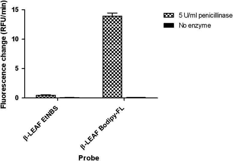
Fig. 8.
Fluorescence change due to the action of penicillinase (-lactamase) on first and second generation probes. 10 μM first and second generation probes were incubated with penicillinase enzyme (final concentration in each reaction) or plain PBS, respectively. The fluorescence change over time was monitored for 60 min and is presented as bar graphs. The -axis shows the measured fluorescence change rate in relative fluorescence units (RFU)/min, which reflects the probe cleavage in respective reactions.

