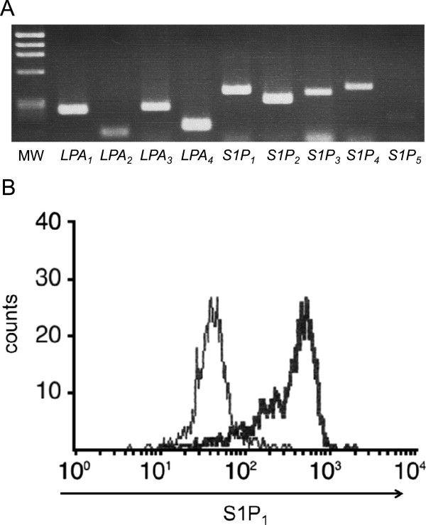
Figure 2.

Expression of S1P and LPA receptors in Jurkat cells. (A) The expression of S1Ps and LPAs in Jurkat cells was examined by RT-PCR. The amplified products for the Jurkat mRNAs for S1P and LPA receptors were resolved in a 2.5% agarose gel. MW indicates the molecular size markers. mRNA transcripts for S1P1, S1P2, S1P3, and S1P4, were found, but that of S1P5 could be hardly detected. Also, LPA1, LPA2, LPA3, and LPA4 were expressed. (B) The surface expression of S1P1 in Jurkat cells was examined by flow-cytometry. Horizontal axis indicates the strength of S1P1 expression as assessed by the indirect FITC fluorescence intensity. The bold line histogram represents staining with anti-S1P1 antibody, while the narrow line represents the negative control. S1P1 was found expressed on Jurkat cells.
