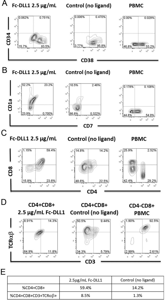
Figure 2. Notch-induced differentiation of CD34+CD38−/low human cord blood HSCs into early T cells.
Flow cytometric analysis of CD34 and CD38 on FSC vs SSC gated cells at day 25 of culture with OP9-DL1 conditioned medium in the presence of immobilized human Fc-DLL1 Notch ligand (2.5 µg/mL) or absence of Notch ligand (negative control). B) Flow cytometric analysis of CD1a and CD7 expression on FSC vs SSC gated cells cultured for 25 days with OP9-DL1 conditioned medium and in the presence of immobilized human Fc-DLL1 Notch ligand (2.5 µg/mL) or absence of Notch ligand (negative control). C) Flow cytometric analysis of CD4 and CD8 expression at day 25 on FSC vs SSC gated cells. PBMC-derived lymphocytes were included in the FACS staining as a positive control for CD4, CD8 and CD3 staining. D) Flow cytometric analysis of CD3 and TCRαβ expression at day 25 on CD4+CD8+ cells cultured in the presence or absence of immobilized Notch ligand (2.5 µg/mL). Isotype staining controls were included in each experiment to determine quadrant gates. E) Table indicating the percentage of the total population that are CD4+CD8+ or CD4+CD8+CD3+TCαβ+.

