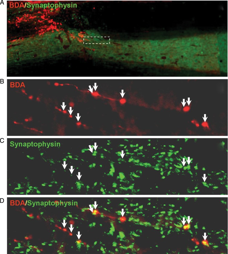
Figure 4.
Cortical delivery of GÖ6976 promotes the synaptic formation of regenerated CST axons. (A) A low-power photomicrograph shows that, in an animal that received cortical delivery of GÖ6976 and lesion site injection of lenti-ChABC after a C4 DH, many BDA-labeled CST axons (red) regenerated beyond the lesion gap (dashed line). (B) High magnification of a boxed area in A shows several regenerated axons labeled with BDA. (C) Distribution of synaptophysin (a presynaptic marker)-IR in the same field presented in B. (D) Merged image of B and C shows the presence of multiple synaptic contacts (arrows) along the lengths of regenerated axons.

