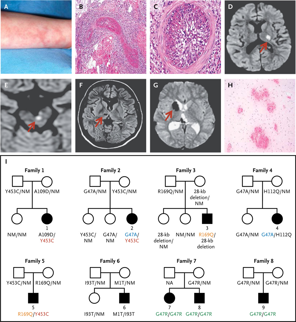
Figure 1. Clinical Findings and Pedigrees of Patients with Deficiency of Adenosine Deaminase 2 (ADA2).
Panel A shows livedo racemosa in Patient 1. Panels B and C show the results of a skin-punch biopsy of the left leg of Patient 4, which revealed vasculitis involving a medium-size blood vessel in the reticular dermis. Panel B shows a dense inflammatory infiltrate with fibrinoid necrosis, and Panel C, the complete occlusion of the vascular lumen (hematoxylin and eosin stain). Panels D through G show the results of brain imaging. Panels D and E show diffusion-weighted axial magnetic resonance images indicating acute small-vessel ischemia in the brain, with Panel D showing ischemia over the left thalamus and posterior limb of the left internal capsule in Patient 5 and Panel E showing ischemia in the left paramedian ventral midbrain in Patient 3; corresponding apparent diffusion coefficient maps are not shown. Panel F shows chronic ischemic changes on axial T1-weighted images in Patient 4 in the form of a small, oval cavitation (lacune) over the right thalamus that resulted from a prior small-vessel occlusion. Panel G shows hemorrhagic events in Patient 2, as detected on T2*-weighted (gradient echo) images. The localized areas of hypointensity over the right caudate head and posterior thalamus correspond to areas of focal accumulation of hemosiderin due to intraparenchymal bleeding. The arrows in Panels D through G point to the area of abnormality. Panel H shows multiple foci of petechial hemorrhages around small-size vessels in the white matter of the brain in Patient 5 (hematoxylin and eosin stain). Panel I shows the pedigrees of the nine affected patients. Patients 1 through 6 were compound heterozygous for eight mutations in CECR1 (cat eye syndrome chromosome region, candidate 1), and Patients 7, 8, and 9 were homozygous for one CECR1 mutation. Squares denote male family members, circles female family members, solid symbols affected family members, and open symbols unaffected family members. Shared CECR1 mutations are color-coded. NM denotes nonmutated.

