Fig. 3.
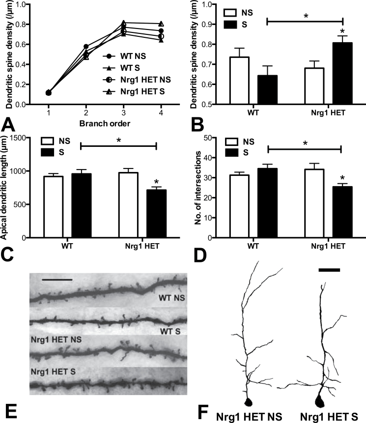
Partial genetic deletion of Nrg1 promoted a repeated stress-induced increase in apical dendritic spine density and a decrease in dendritic lengths and complexity in layers II/III of the medial prefrontal cortex (mPFC). (A) Dendritic spine density per µm of apical dendrites with increasing dendritic branch order. (B) Dendritic spine density on fourth order dendrites. (C) Cumulative apical dendritic lengths and (D) no. of intersections as determined by Sholl analysis. Repeatedly stressed Nrg1 HET mice exhibited significantly increased dendritic spine density and decreased dendritic lengths and dendritic complexity, in the mPFC compared with nonstressed Nrg1 HET mice or stressed WT mice, *Ps < .05. (E) Representative photomicrographs of Golgi-stained dendritic spines on fourth order apical dendrites in the mPFC, scale bar = 5 µm. (F) Representative tracings of Golgi-stained mPFC apical dendrites in nonstressed vs stressed Nrg1 HET mice, scale bar = 50 µm. WT, wild-type mice; Nrg1 HET, neuregulin 1 heterozygous mice; NS, nonstressed homecage controls; S, mice subjected to restraint stress.

