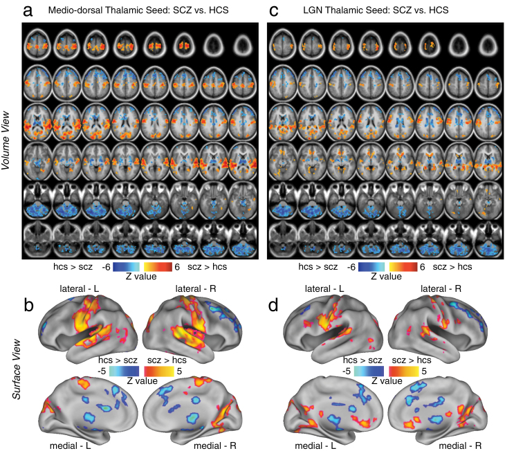
Fig. 1.
Differences in mediodorsal (MD) vs occipital-projecting thalamic nuclei in schizophrenia (SCZ). Threshold-free cluster enhancement36 whole-brain corrected volume and surface maps of group differences between SCZ patients and matched healthy comparison subjects (HCS). Results are shown for the MD prefrontal-projecting thalamic seed (panels a and b) and the lateral geniculate nucleus (LGN) occipital-projecting seed (panel b and c), both defined using the FSL atlas19 (see “Method” section for a detailed description of seed selection). Red foci mark regions where SCZ showed statistically higher connectivity than HCS, whereas blue foci show regions where SCZ show statistically lower connectivity than HCS for a given thalamic seed. For a complete list of regions and statistics see table 2. Note: Panels a and c show the results in a volume representation, whereas panels b and d show the same data mapped onto a surface representation.

